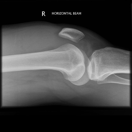

Calcium pyrophosphate crystals mean you have pseudogout.īacteria in the joint fluid that are causing an infection may be seen under a microscope after being colored with a Gram stain (a special dye). Uric acid crystals in the joint mean you have gout. Large numbers of white blood cells may be caused by gout, pseudogout, other types of arthritis (such as rheumatoid arthritis), psoriatic arthritis, injury, or infection. Large numbers of red blood cells may be caused by bleeding in the joint from injury, inflammation, or abnormal clotting of the blood. Milky white may be caused by infection or inflammation or a condition such as gout. Osteoarthritis simply means that the protective cartilage of the knee has worn out. This is normally caused by an injury and the knee swelling comes on rapidly (within minutes). The most common reason for knee replacement surgery is Osteoarthritis. A deep, dark red color may be caused by bleeding in the joint. Why Does The Knee Swell Bleeding in the Joint: aka Haemarthrosis. Slightly cloudy fluid may be caused by inflammation, gout, or pseudogout. No bacteria are seen, and no organisms grow in the culture.īacteria are seen, or organisms grow in the culture. Large numbers of red or white blood cellsĬrystals (seen under a special microscope with polarized light) No large numbers of red or white blood cells The results from a culture usually take a few days. Sometimes the excess blood ends up in the synovial fluid. Hemophilia is an inherited disorder that can cause excessive bleeding. Infection in a joint Bleeding disorder, such as hemophilia. The results of a joint fluid analysis are usually ready the same day. When it affects the knee, it may be referred to as knee effusion or fluid on the knee. An elastic bandage may also be wrapped around your joint, such as your knee, to reduce swelling. It can help keep fluid from building up again.Ī tight (pressure) bandage will be placed over the site to reduce swelling and bruising. A cortisone shot may be given into the joint before the needle is removed. Samples of the fluid may be put in special tubes or containers and sent to the lab. A syringe attached to the needle is used to remove a sample of joint fluid. For young children, a sedative may also be given.Ī long, thin needle is slowly inserted in the joint area.
/kneepainmedreview-01-5c7d9f26c9e77c0001fd5a7d.png)
BLOODY FLUID IN KNEE JOINT SKIN
A local anesthetic is often injected into the skin over the joint. The skin over the joint area will be cleaned with antiseptic solution. Your doctor may use ultrasound to guide the needle placement.

Your doctor will examine the joint to find out where the needle should be inserted. Blood tests : Your provider may have you undergo blood tests to see if your body’s immune system is responding to an infection and/or to rule out. You will sit or lie down on an examining table. Synovial fluid aspiration: Your healthcare provider may withdraw fluid from your affected joint with a fine needle to check it for bacteria. Depending on which joint will be examined, you may be asked to undress and put on a hospital gown. Joint fluid analysis can be done in your doctor's office, clinic, operating room, or emergency room.


 0 kommentar(er)
